Keratinocyte Lifecycle
Keratinocytes are the predominant cell type in the epidermis and form the structural foundation of the skin’s barrier. Their lifecycle—from stem cell to cornified cell—is a meticulously regulated process that renews the epidermis approximately every 28 days in healthy adults. This cycle ensures continuous repair from daily wear, maintains hydration, protects against pathogens, and enables rapid response to injury. Understanding keratinocyte differentiation provides a crucial framework for interpreting wound healing, dermatological disease, and the physiological basis of the skin barrier.
What You Need to Know
Keratinocytes are the dominant cell type of the epidermis and are responsible for forming the skin’s physical and biochemical barrier. They originate from stem cells in the basal layer, where continuous cell division replenishes the epidermis. From this proliferative zone, keratinocytes migrate upward through the epidermal layers, undergoing a tightly regulated process of differentiation that transforms them from living cells into highly specialised barrier components.
As keratinocytes move through the epidermis, several coordinated changes occur:
Progressive accumulation and reorganisation of keratin filaments
Synthesis and secretion of lipids essential for barrier formation
Loss of nuclei and organelles during terminal differentiation
Formation of a rigid protein envelope that strengthens the cell surface
By the time keratinocytes reach the stratum corneum, they have become flattened, non-viable corneocytes embedded within a lipid-rich extracellular matrix. Together, these corneocytes form a resilient, water-resistant layer that provides mechanical strength and limits transepidermal water loss. The final stage of the lifecycle is desquamation, where corneocytes are shed from the surface in a controlled manner. This balance between cell production, differentiation, and shedding is essential for maintaining epidermal thickness, barrier integrity, and effective protection against environmental injury.
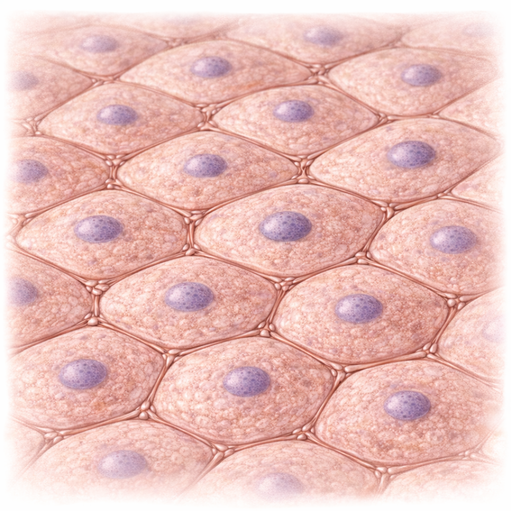
Image: Keratinocytes within the epidermis form a tightly connected network, linked by desmosomes to provide structural strength and maintain the integrity of the skin barrier.
Beyond the Basics
Basal layer and stem cell proliferation
Keratinocyte renewal begins in the stratum basale, a single layer of proliferative stem cells anchored to the basement membrane through hemidesmosomes. These attachments provide mechanical stability and also allow basal keratinocytes to sense signals from the underlying dermis. Continuous mitotic activity within this layer replenishes the epidermis and replaces corneocytes lost at the surface. Proliferation is tightly regulated by growth factors such as epidermal growth factor and transforming growth factor alpha, and is further influenced by hormones, mechanical stress, injury, and inflammatory mediators.
Basal keratinocytes predominantly express keratins 5 and 14, which form intermediate filaments that provide resistance to mechanical stress at the dermoepidermal junction. As daughter cells detach from the basement membrane, they lose direct access to dermal signalling and begin the process of terminal differentiation. This upward migration marks the transition from a proliferative to a differentiating cell state and initiates the orderly progression through the epidermal layers.
Stratum spinosum and cytoskeletal reinforcement
In the stratum spinosum, keratinocytes increase their mechanical connectivity through the formation of numerous desmosomes. These intercellular junctions link adjacent cells, distributing mechanical forces across the epidermis and preventing tissue separation during shear or stretch. The prominent appearance of these junctions under microscopy gives the layer its characteristic spiny appearance.
During this stage, keratin expression shifts from basal keratins to keratins 1 and 10, producing a stronger and more resilient cytoskeleton. Keratinocytes in the stratum spinosum remain metabolically active and contribute to barrier preparation by synthesising lipids, antimicrobial peptides, and structural proteins. This layer also contains Langerhans cells, which provide immune surveillance and integrate antigen detection with ongoing epidermal renewal and repair.
Stratum granulosum and barrier assembly
As keratinocytes enter the stratum granulosum, differentiation accelerates and profound biochemical changes occur. Cells accumulate keratohyalin granules containing profilaggrin, which is later cleaved into filaggrin. Filaggrin aggregates keratin filaments into dense bundles, transforming the cytoplasm into a compact, mechanically resistant structure.
At the same time, lamellar bodies within keratinocytes secrete lipids, including ceramides, cholesterol, and free fatty acids, into the extracellular space. These lipids organise into lamellar sheets that form the water-resistant barrier of the stratum corneum. Nuclear and organelle degradation begins during this stage, converting the cell from a living unit into a structural component optimised for barrier function.
Stratum corneum and cornification
The stratum corneum consists of multiple layers of flattened, non-viable corneocytes embedded in a lipid-rich matrix. Each corneocyte is surrounded by a cornified envelope composed of cross-linked proteins and bound lipids, creating a structure with remarkable resistance to mechanical stress, chemical exposure, and microbial invasion. This layer represents the final product of keratinocyte differentiation.
Breakdown products of filaggrin within corneocytes form natural moisturising factors that attract and retain water, supporting hydration and maintaining the acidic surface environment of the skin. Through this combination of physical structure, lipid organisation, and biochemical buffering, the stratum corneum serves as the primary barrier against transepidermal water loss and environmental injury.
Desquamation and controlled shedding
The keratinocyte lifecycle concludes with desquamation, the controlled shedding of corneocytes from the skin surface. This process depends on enzymatic degradation of corneodesmosomes by proteases such as kallikreins. Enzyme activity is tightly regulated by pH, natural moisturising factors, and protease inhibitors to ensure gradual and even shedding.
When this regulatory balance is disrupted, abnormal scaling can occur. Excessive desquamation leads to dry, flaky skin, while impaired shedding results in corneocyte retention and thickened scales. Proper regulation of desquamation is therefore essential for maintaining surface smoothness, barrier integrity, and epidermal homeostasis.
Regulation of epidermal turnover
In healthy adult skin, complete epidermal turnover takes approximately four weeks, although this timeframe varies across the lifespan and in disease states. Turnover is faster in infancy, slows with ageing, and becomes markedly accelerated in conditions such as psoriasis, where differentiation is incomplete and barrier formation is impaired.
Hormonal influences, nutritional status, circadian rhythms, mechanical loading, and systemic illness all modulate keratinocyte proliferation and differentiation. This dynamic regulation allows the epidermis to adapt to injury and environmental stress while preserving structural integrity. Disruption at any stage of the keratinocyte lifecycle can compromise barrier function and underlie a wide range of dermatological conditions.
Clinical Connections
Many common skin conditions arise from disruption at specific stages of the keratinocyte lifecycle. When proliferation, differentiation, or desquamation becomes unbalanced, the epidermis loses its ability to maintain an effective barrier, regulate hydration, and repair itself efficiently. Clinical presentations often reflect where in the lifecycle the disruption is occurring.
In practice, altered keratinocyte turnover tends to present in a small number of recognisable patterns:
Excessive proliferation with incomplete differentiation
Impaired barrier formation due to defective protein or lipid processing
Reduced regenerative capacity following cellular injury
Psoriasis is a clear example of accelerated keratinocyte turnover. Cells proliferate rapidly in the basal layer and reach the skin surface prematurely, resulting in thickened plaques with incomplete cornification and excessive scaling. Atopic dermatitis reflects a different disruption, where defective filaggrin processing and impaired lipid organisation weaken the stratum corneum. This leads to increased water loss, heightened sensitivity to irritants, and greater susceptibility to inflammation and infection.
Ageing alters the keratinocyte lifecycle by slowing proliferation and reducing lipid and natural moisturising factor production. These changes result in dry, fragile skin with delayed wound healing and reduced tolerance to mechanical stress. Similar effects are seen when mitotic activity in the basal layer is disrupted by chemotherapy, radiation therapy, or severe systemic illness. In these settings, epidermal thinning, ulceration, and increased infection risk reflect impaired renewal rather than primary barrier disease.
Understanding the keratinocyte lifecycle supports informed clinical decision-making in wound management and skin care. Knowledge of turnover rates, barrier formation, and desquamation helps guide dressing selection, moisturisation strategies, and early recognition of conditions where epidermal repair is compromised. Linking visible skin changes back to underlying cellular processes allows more accurate assessment and targeted intervention across a wide range of clinical contexts.
Concept Check
What key structural and biochemical changes occur as keratinocytes move from the basal layer to the stratum corneum?
How does filaggrin contribute to both the barrier function and hydration of the skin?
Why is desquamation essential for maintaining a healthy epidermis?
How does the lipid matrix in the stratum corneum support barrier integrity?
What factors influence the rate of epidermal turnover?