Protective Structures of the CNS: The Meninges, Cerebrospinal Fluid & Blood–Brain Barrier
The central nervous system is highly specialised, irreplaceable tissue that must be protected from mechanical injury, infection, and fluctuations in the internal environment. Unlike many other tissues, neurons have limited regenerative capacity, making protection essential for survival and long-term function. To achieve this, the brain and spinal cord are safeguarded by three integrated protective systems: the meninges, cerebrospinal fluid (CSF), and the blood–brain barrier (BBB). These structures not only provide physical protection but also regulate pressure, nourish neural tissue, remove waste, and strictly control which substances can enter the neural environment. Dysfunction in any of these protective systems can rapidly compromise neurological function and become life-threatening.
What You Need to Know
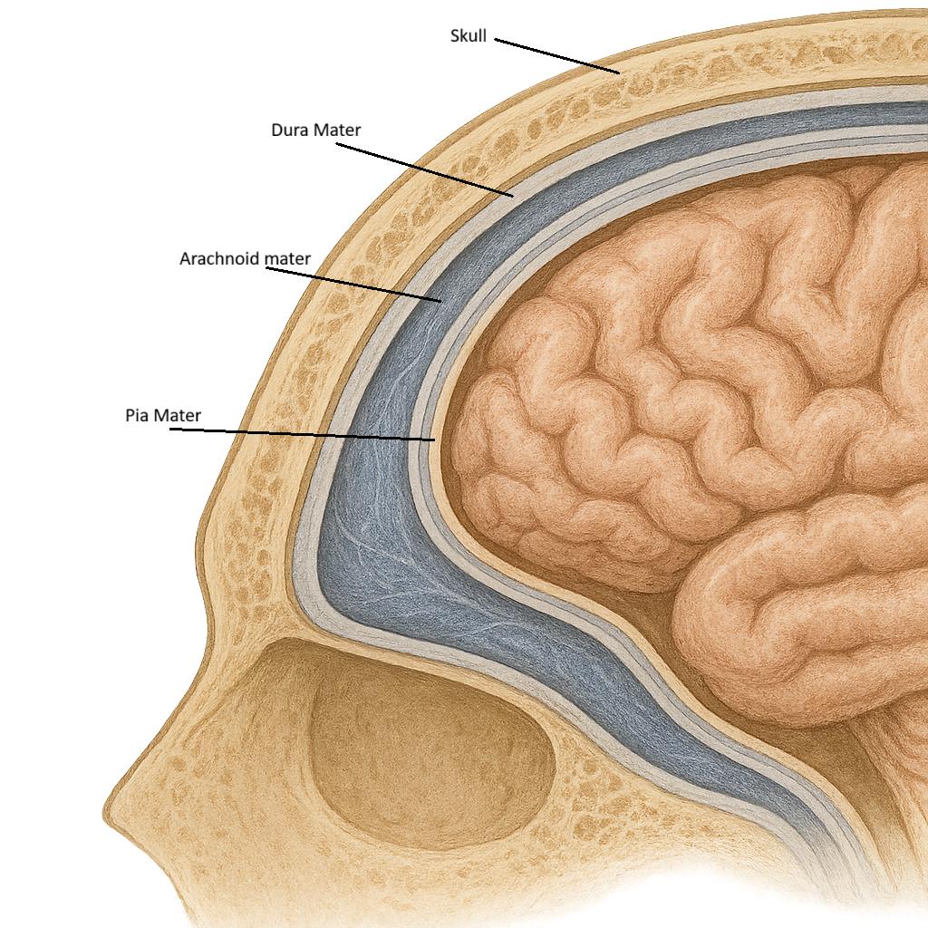
The meninges are three connective tissue layers that surround the brain and spinal cord. The outermost layer is the dura mater, a tough, fibrous membrane that provides strong mechanical protection and forms venous sinuses for blood drainage. Beneath the dura is the arachnoid mater, a delicate, web-like layer that creates a cushioning space for cerebrospinal fluid. The innermost layer is the pia mater, a thin, highly vascular membrane that tightly adheres to the surface of the brain and spinal cord, following every sulcus and gyrus.
Key features of the meninges:
Dura mater: tough outer protection and venous drainage
Arachnoid mater: creates space for CSF cushioning
Pia mater: closely adheres to and nourishes neural tissue
Subarachnoid space: contains CSF and major blood vessels
Between the arachnoid and pia mater lies the subarachnoid space, which contains cerebrospinal fluid and major cerebral blood vessels. This space allows CSF to circulate freely around the CNS and absorb mechanical shock.
Cerebrospinal fluid (CSF) is a clear, colourless fluid produced by the choroid plexuses within the ventricles of the brain. It circulates through the ventricular system, into the subarachnoid space, and around the spinal cord before being reabsorbed into the venous circulation via arachnoid granulations.
The blood–brain barrier (BBB) is a highly selective barrier formed by tightly packed endothelial cells lining cerebral capillaries, supported by astrocytes and pericytes. It strictly controls which substances can pass from the bloodstream into the brain’s extracellular fluid.
Beyond the Basics
The Meninges as a Mechanical and Metabolic System
The meninges form a continuous, layered system that provides both mechanical stability and physiological support to the central nervous system. Each layer contributes in a distinct but integrated way.
The dura mater forms a tough outer shell that anchors the brain within the skull. Its inward folds, including the falx cerebri and tentorium cerebelli, divide the cranial cavity into compartments that limit brain displacement during rapid head movement. These dural reflections also create channels for venous drainage, linking structural support to vascular regulation.
Beneath the dura, the arachnoid mater creates a sealed compartment that holds cerebrospinal fluid. This layer functions as a hydraulic suspension system, allowing the brain to float rather than rest on the skull base. This arrangement distributes mechanical forces evenly and prevents focal pressure on delicate neural tissue.
The pia mater closely follows the contours of the brain and spinal cord, investing every sulcus and fissure. It supports fine blood vessels as they penetrate neural tissue and facilitates the exchange of nutrients and metabolic waste between the circulation and neurons.
Together, the meninges transform the brain from a vulnerable soft tissue into a mechanically stabilised, metabolically supported organ.
Cerebrospinal Fluid as a Dynamic Regulatory Medium
Cerebrospinal fluid is not simply a protective cushion — it is an actively regulated internal environment for the central nervous system. CSF is continuously produced by the choroid plexus and circulates through the ventricles and subarachnoid space before being reabsorbed into the venous system.
This constant turnover serves several critical functions. By surrounding the brain and spinal cord, CSF provides buoyancy, reducing the effective weight of the brain from approximately 1.4 kg to less than 50 g. This prevents compression of neural structures at the base of the skull and spinal canal.
CSF also acts as a shock absorber, dispersing kinetic energy during movement or impact. In addition, it contributes to chemical homeostasis by distributing nutrients, removing metabolic waste, and stabilising the ionic environment required for precise electrical signalling. Because CSF volume and flow are tightly regulated, it also plays a central role in maintaining intracranial pressure within narrow physiological limits.
The Blood–Brain Barrier as a Selective Interface
The blood–brain barrier (BBB) is a specialised vascular interface that separates the circulating blood from the neural environment. It is formed by capillary endothelial cells joined by tight junctions, supported by astrocyte end-feet and a basement membrane. This structure eliminates the gaps present in most capillaries, preventing uncontrolled movement of substances into the brain.
The BBB allows lipid-soluble molecules such as oxygen, carbon dioxide, and certain drugs to diffuse freely, while essential nutrients like glucose and amino acids are transported via specific carrier proteins. Most ions, large molecules, proteins, and immune cells are excluded unless specialised transport or signalling mechanisms are activated.
This selective permeability ensures that neurons operate within a stable chemical environment, protected from fluctuations in blood composition, circulating toxins, and inflammatory mediators. Such stability is critical for synaptic transmission, action potential generation, and neural network function.
Integration of Barriers and Neural Protection
The meninges, cerebrospinal fluid, and blood–brain barrier form a coordinated protective system rather than independent structures. The meninges provide mechanical support, CSF provides hydraulic and chemical buffering, and the BBB provides molecular regulation.
Together, they create an environment in which neurons can function with extraordinary precision, shielded from mechanical stress, chemical instability, and biological threats. This layered system of protection is one of the defining features of the central nervous system and underpins the brain’s ability to sustain complex, high-speed signalling over a lifetime.
Clinical Connections
Disorders of the meninges and cerebrospinal fluid typically present with recognisable clinical patterns. Meningitis reflects inflammation within the subarachnoid space, producing headache, photophobia, fever, neck stiffness, and, in more severe cases, altered consciousness due to rising intracranial pressure and impaired cerebral perfusion.
Traumatic bleeding between meningeal layers produces distinct syndromes depending on location. Epidural haematomas, usually due to arterial bleeding (classically the middle meningeal artery), can deteriorate rapidly following a brief lucid period. Subdural haematomas, typically venous, often present more gradually with confusion, headache, or reduced consciousness. Both increase intracranial pressure and compress underlying brain tissue. In clinical practice, these conditions are often distinguished by patterns such as:
Epidural haematoma: head trauma followed by a brief loss of consciousness, a lucid interval, then rapid neurological deterioration
Subdural haematoma: head trauma, often minor, with gradual onset confusion or reduced level of consciousness, particularly in older adults
Subarachnoid haemorrhage: sudden, severe “thunderclap” headache, often described as the worst headache of life, with possible neck stiffness, photophobia, and reduced consciousness
Meningitis: fever, neck stiffness, photophobia, and altered mental state, sometimes accompanied by a rash in meningococcal disease
Raised intracranial pressure: worsening headache, vomiting, reduced level of consciousness, and possible pupillary changes
Hydrocephalus in adults: gait disturbance, cognitive decline, and urinary incontinence
Abnormal cerebrospinal fluid dynamics further highlight the importance of pressure regulation. Hydrocephalus results in ventricular enlargement and progressive neurological dysfunction, with presentation varying by age. Lumbar puncture remains essential for diagnosing infection, subarachnoid haemorrhage, and inflammatory conditions, but is contraindicated in suspected raised intracranial pressure due to the risk of herniation.
Disruption of the blood–brain barrier contributes to cerebral oedema and secondary injury in conditions such as stroke, trauma, infection, and multiple sclerosis, and also determines which medications can effectively act within the central nervous system.
Concept Check
Why does meningeal inflammation cause neck stiffness and photophobia?
How does cerebrospinal fluid protect the brain mechanically and chemically?
Why does hydrocephalus increase intracranial pressure?
How does the blood–brain barrier protect neural tissue from toxins?
Why does BBB disruption worsen outcomes after stroke and trauma?