PERICARDITIS: Inflammation of the Pericardium
Pericarditis is inflammation of the pericardial layers, the fibrous parietal pericardium and the serous visceral pericardium (epicardium). It disrupts the normally smooth, frictionless environment surrounding the heart and may lead to chest pain, pericardial effusion and, in severe cases, cardiac tamponade.
Pericarditis can occur as an isolated condition or as part of a broader systemic process. Viral infections are the most common cause, but autoimmune disorders, post-myocardial infarction inflammation, uraemia and malignancy are also important contributors. The clinical course varies widely: some cases resolve quickly, whereas others progress to recurrent or chronic constrictive pericarditis.
What You Need to Know
Pericarditis is inflammation of the pericardium, the double-layered sac that surrounds the heart. Under normal conditions, a thin layer of lubricating fluid within the pericardial space reduces friction as the heart contracts and relaxes. When inflammation develops, vascular permeability increases and proteins, inflammatory cells, and fluid accumulate within the pericardial cavity. The inflamed pericardial layers may become roughened, and fibrin deposition can occur, contributing to the characteristic chest pain and audible friction rub.
The most common symptom is sharp, pleuritic chest pain that is typically worse with deep inspiration or when lying flat, and improves when sitting upright or leaning forward. This positional nature of pain helps distinguish pericarditis from myocardial ischaemia. A pericardial friction rub, described as a scratchy or high-pitched sound heard best along the left sternal border, is a key clinical sign caused by inflamed pericardial surfaces rubbing together. Inflammatory processes may be triggered by viral infection, autoimmune disease, post-myocardial infarction syndromes, uraemia, malignancy, or following cardiac surgery.
The main complications arise from fluid accumulation or chronic inflammation:
Pericardial effusion, where excess fluid collects in the pericardial space.
Cardiac tamponade, when rising pericardial pressure restricts ventricular filling and reduces cardiac output.
Constrictive pericarditis, a chronic condition where fibrosis and thickening limit normal cardiac expansion.
Most cases are self-limiting with appropriate anti-inflammatory treatment, but close monitoring is important because rapid fluid accumulation can compromise haemodynamics even when the total volume of fluid is modest.
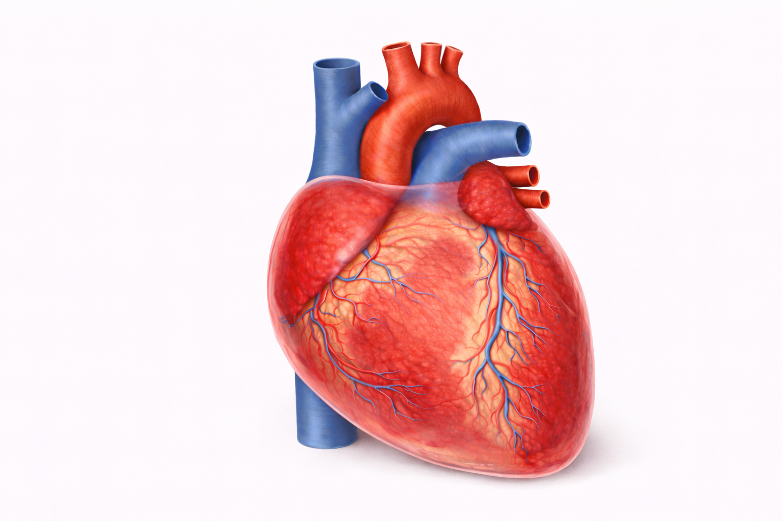
Image: In pericarditis, inflammation of the pericardium causes the normally thin, transparent sac to become thickened, reddened, and irritated around the surface of the heart.
Beyond the Basics
Mechanisms of Inflammation and Fluid Accumulation
Pericarditis begins with inflammation of the visceral and parietal pericardial layers. Inflammatory mediators increase vascular permeability within pericardial capillaries, allowing plasma proteins and leukocytes to leak into the pericardial space. The resulting fluid is typically an exudate, meaning it is protein-rich and driven by inflammation rather than simple pressure imbalance. Depending on the underlying cause, the effusion may be serous, fibrinous, purulent, or haemorrhagic.
Fibrinous pericarditis is characterised by deposition of fibrin strands along the inflamed surfaces, producing the classic scratchy friction rub heard on auscultation. Purulent pericarditis reflects bacterial infection and involves thick, neutrophil-rich fluid that can rapidly compromise haemodynamics and carries high morbidity. Haemorrhagic effusions may occur in malignancy, tuberculosis, or after cardiac injury. The rate of fluid accumulation is clinically crucial. When fluid collects slowly, the pericardium can stretch over time and accommodate large volumes with relatively minor changes in pressure. In contrast, rapid accumulation, even in modest volumes, can sharply elevate intrapericardial pressure and impair cardiac filling.
Haemodynamic Consequences: From Effusion to Tamponade
As fluid accumulates, intrapericardial pressure gradually rises. Once this pressure exceeds the normal filling pressures of the heart, diastolic expansion becomes restricted. The right atrium and right ventricle are typically affected first because they operate at lower pressures than the left-sided chambers. Reduced diastolic filling leads to decreased stroke volume and a compensatory increase in heart rate to maintain cardiac output.
Cardiac tamponade develops when intrapericardial pressure critically limits ventricular filling throughout diastole. Venous return is impeded, and cardiac output falls sharply. Clinically, tamponade presents with hypotension, raised jugular venous pressure, and pulsus paradoxus, which is an exaggerated fall in systolic blood pressure during inspiration. Without urgent intervention, such as pericardiocentesis to drain the fluid, circulatory collapse may occur. The severity of tamponade depends more on the speed of fluid accumulation than the absolute volume present.
Post-Myocardial Infarction and Autoimmune Pericarditis
Pericarditis may complicate myocardial infarction in two distinct forms. Early post-infarction pericarditis occurs within the first few days and results from local extension of inflammation from the necrotic myocardium to the adjacent pericardium. Dressler’s syndrome develops weeks later and is immune-mediated, likely triggered by an autoimmune response to myocardial antigens exposed during infarction. It is characterised by fever, pleuritic chest pain, and often large pericardial effusions.
Autoimmune conditions such as systemic lupus erythematosus and rheumatoid arthritis can also involve the pericardium through chronic immune activation and immune complex deposition. In these cases, pericarditis may recur or become persistent, reflecting ongoing systemic inflammation rather than an isolated cardiac process.
Constrictive Pericarditis
Chronic or recurrent inflammation may lead to progressive fibrosis, thickening, and sometimes calcification of the pericardial layers. The once-compliant pericardial sac becomes rigid and non-distensible, restricting normal ventricular filling during diastole. Unlike tamponade, where fluid creates external pressure, constrictive pericarditis is caused by a stiff fibrotic shell encasing the heart.
The haemodynamic pattern resembles right-sided heart failure, with elevated venous pressures, peripheral oedema, ascites, and reduced cardiac output. Because the restriction is mechanical and chronic, medical therapy is usually insufficient once advanced fibrosis has developed. Definitive management often requires surgical pericardiectomy, where the thickened pericardium is removed to restore ventricular filling capacity.
Clinical Connections
Pericarditis is primarily a clinical diagnosis supported by characteristic findings. Sharp, pleuritic chest pain that improves when sitting forward is a key feature, and the presence of a pericardial friction rub on auscultation strongly supports the diagnosis. Electrocardiography typically demonstrates diffuse ST-segment elevation with PR-segment depression, changes that reflect widespread epicardial inflammation rather than regional myocardial ischaemia. Unlike myocardial infarction, these ST elevations are usually concave and occur across multiple leads. Inflammatory markers such as C-reactive protein and erythrocyte sedimentation rate are often elevated and can help track disease activity over time.
Several findings help identify complications or alternative diagnoses:
Large or rapidly accumulating pericardial effusion may present with tachycardia, hypotension, and rising jugular venous pressure.
Persistent fever or purulent features raise concern for bacterial infection.
Recurrent chest pain with elevated inflammatory markers suggests relapsing or inadequately treated inflammation.
Echocardiography is essential to assess for pericardial effusion and to evaluate for haemodynamic compromise. In tamponade, imaging may demonstrate right atrial or right ventricular diastolic collapse, reflecting elevated intrapericardial pressure that impairs filling. Treatment typically includes nonsteroidal anti-inflammatory drugs to reduce inflammation and pain, with colchicine added to decrease recurrence risk. Glucocorticoids may be used when symptoms are refractory or when autoimmune disease is the underlying driver, though they are generally reserved for selected cases because of recurrence risk. Close monitoring is necessary to detect early signs of tamponade, including tachycardia, narrowing pulse pressure, hypotension, or escalating jugular venous distension. Education focuses on gradual return to activity once symptoms and inflammatory markers improve, adherence to anti-inflammatory therapy, and prompt review if chest pain or breathlessness worsens.
Concept Check
Why does pericarditis produce pleuritic chest pain that improves when sitting forward?
How can inflammation of the pericardium lead to pericardial effusion?
Why does rapid pericardial fluid accumulation cause tamponade more readily than slow accumulation?
What ECG pattern is typical of acute pericarditis?
What differentiates constrictive pericarditis from pericardial tamponade?